Simulated data for convex array
The RF data was obtained from simulating wires in a phantom
by using the Field
II program and a convex array probe. The geometry of the
phantom is shown below, where wires are placed with a spacing of
1 cm.
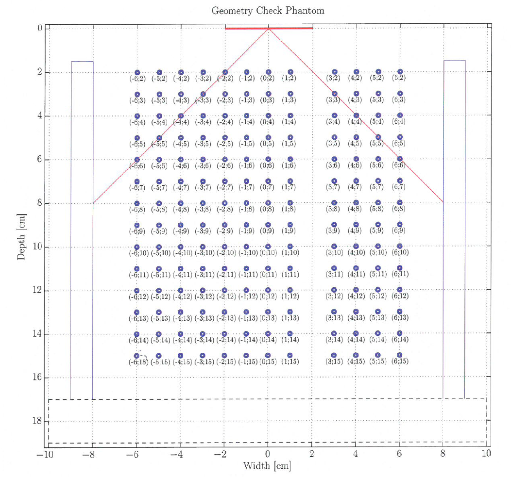
Geometry of the matrix wire phantom
| Scan mode: | B-mode (Duplex mode) |
| Transducer: | BK 8820e, 3.5 MHz convex array probe |
| Number of elements | 192 |
| Transducer center frequency | 3.50 MHz |
| Number of active elements in transmit | 64 |
| Height of one element | 13 mm |
| Width of one element | 0.30 mm |
| Kerf (gap) between elements | 0.03 mm |
| Electronic focus depth | 42.2 mm |
| Convex radius | 60.3 mm |
| Elevation focus | 65 mm |
| B-mode frame rate: | 0.78 f/s |
| Speed of sound: | 1491 m/s |
| Pulse repetition frequency: | 100 Hz |
| Sampling frequency: | 17.5 MHz |
| Sample format: | double |
The data is stored in the zip archive (642 MBytes):
https://courses.healthtech.dtu.dk/22485/files/ult_data/simulation/phantom_matrix/
It is taken from a duplex sequence with intermixed B-mode and duplex flow emissions.
The B-mode data are in the directory: duplex/B_mode/seq_0001, where seq_0001 holds all
the data for the first image. A file elem_data_em0XXX.mat exist for each emission, where XXX
is from 1 to 129. For the first file elements 1 to 64 has been used for transmission and
for the next element 2 to 65 and so forth. Element 1 has the most negative x coordinate value.
The file holds the matix samples with the received signals for all 192 transducer elements.
The zip file also hold the files parameters.mat
and sim_parameters.mat, which hold structures with all the variables used for the simulation
of the phantom.
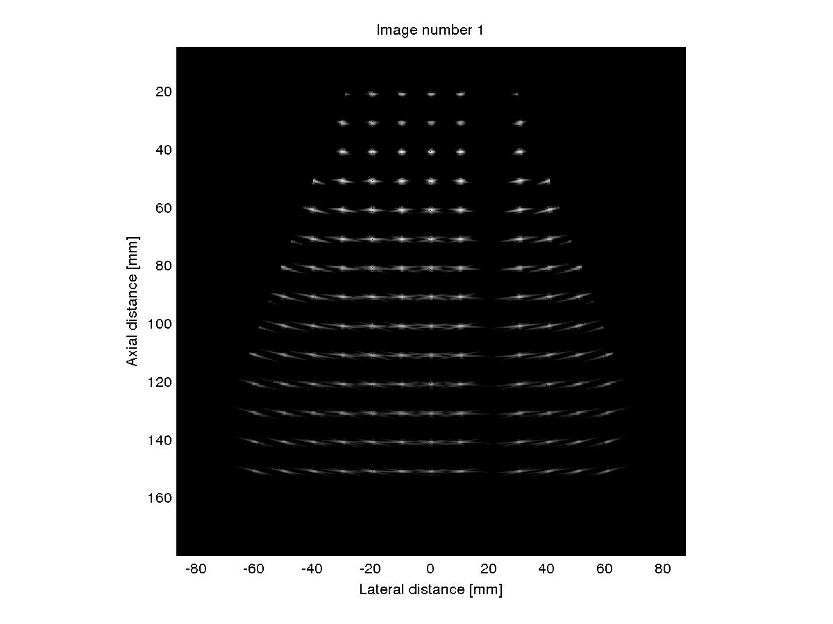
Simulated image for the matrix wire phantom
| 

 Print Version