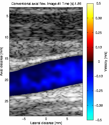
In-vivo color flow mapping data from carotid artery
The data were acquired with the experimental scanner RASMUS at the time of
peak systole of a healthy 30 year old male. The transducer was a B-K 8812
linear array transducer.
The parameters for the measurement are given below:
| Ultrasound scanner: | RASMUS experimental ultrasound scanner |
| Scan mode: | CFM and B-mode |
| Transducer: | B-K 8812, 6.2 MHz linear array probe |
| Matlab file name: | cfm_carotis.mat |
| Sampling frequency: | 40 MHz |
| Resolution: | 16 bits samples |
| | |
Speed of sound c: | 1540 m/s |
| Puls repetition frequency fprf | 6 kHz |
| Center frequency f0 | 5 MHz |
| Focus depth: | 18 mm |
| Cycles in pulse: | 8 |
| Start depth of data: | 1 mm |
| End depth of data: | 30 mm |
| Dimension left to right: | x_min: -9.75 mm to x_max 9.75 mm |
The data can be obtained from the ftp-server from the directory:
https://courses.healthtech.dtu.dk/22485/files/ult_data/in-vivo/cfm_carotis/
in the file
cfm_carotis.mat. The size is 3.7 Mbytes.
The data can be loaded into Matlab. rf_cfm_data is the variable containing the
data for velocity estimation. It is a three dimensional matrix. The first index
is the sample number, the second is the emission number (1-64) and the third
is the position in the image (1-16) equally spaced out between x_min and x_max.
The 16 velocity lines covers the whole area scanned by the B-mode image. The
data are stored as 16 bits signed integers to save space.
bmode_data contains data for the B-mode image.
D is a discriminator matrix to seperate the blood from the tissue.
It has a value of 1 where the velocity should be shown in the CFM image
and the value 0 where no blood is present.
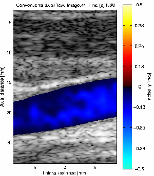
The final image made from the data should look as above.
| 

 Print Version