CT data
On this page you can gain access to a database of CT images and projection
data for the images, that can be used for making reconstruction algorithms.
Data for some phantom objects and various programs for artificial phantoms
and data projections are also given here.
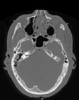
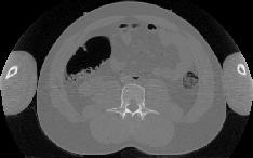
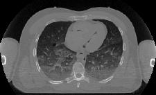
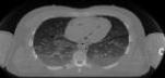
The clinical images shown on these pages have been obtained from
the
Visible Human Project. They were created from scanning a human cadaver
with CT and MRI scanners and then subsequently slicing the cadaver into
1 mm sections for taking photographs.
A further description of the data can be found at
here
and a description of the program can be found
here.
You can get to the different data pages by clicking on one of the images
or the text below them.
A slide showing Housfiled units for a CT scan can be donwloaded
here. The slide
is taken from: E. Krestel (ed.): Imaging systems for medical
diagnostics, Siemens, 1990.
A table of Mass Attenuation Coefficients
and Mass Energy-Absorption Coefficients can be
found at the following reference:
http://www.physics.nist.gov/PhysRefData/XrayMassCoef/cover.html
A radiologic image atlas can be found at:
http://rad.usuhs.mil/rad/iong/homepage.html
| 

 Print Version