Assignment 1: Estimation of T1 and relative PD from 3D-FLASH
Please conduct and describe T1 and relative PD estimation from a series of 3D FLASH images acquired with different tip angles. The practical part is discussed on the course homepage under "Data acquisition". The analysis was discussed in connection with exercises in prior weeks. Use the software developed and validated during these.
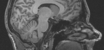
Supply an example of calculated T1 and relative PD images in coronal, sagital and transversal orientations, and comment on these. Describe the variation between different tissue types. Also compare values between similar tissues at different locations, particularly white matter in corpus callosum and gray matter in the putamen. Compare to approximate general values found in Nishimura or other literature (deviations are expected). Discuss relevant confounds and error sources for the specific data sets.
Document the acquisition, the analysis and the findings in a minimal, but informative form as described on the course homepage under "Assignments". Include central formulas and definitions. Please also supply the used software as an appendix so useful feedback can be provided. The style of programming or of commenting will not be evaluated. Advice is below, in Learn/Content/ReportInfo, and on the course homepage (different kinds of advice the 3 places). A message is sent via DTU Learn if there are significant changes to the texts.
Important advice:
As written elsewhere the reports are strictly individual or written in groups of 2 (handing in exactly the same report), but you can work with anybody for the analysis (specify group members as comments in the source code, if you do). Supply the report in PDF format. Keep the actual text at maximally 3 pages including formatting elements (if 11pt font size). Any amount of graphs, formulas and tables can be inserted in the text (it does not count wrt. the page limit).
You are not required to use "your own" data, but please use data from this year for the hand-in, if possible.
It is needed to have data with a -common scaling of the three datasets. You should check the scaling of the recorded data, but not document it in the report. The following matlab visualization of slice 80 should generally reflect that the signal is largest near the tissue-dependent Ernst angle:
imagesc([i1.img(:,:,80), i2.img(:,:,80), i3.img(:,:,80)])
Choose a slice that shows much brain. In particular, the acquisition with small tip angle (~5 or smaller) should appear relatively dark if TR is around 10 ms. If not, ask about rescaling or use another method of reading in data. Use data on DTU Learn until it is resolved.
Remember that only 10 minutes of correction time is allocated per report for the initial submission (see homepage), which is completely inadequate if the report is not well written. You can upload a corrected report a few weeks before the exam. Only this wil be used for grading. Do not get inspiration from earlier reports - I hate reporting potential fraud.
| 
 Print Version