Description of 22481 Introduction to medical imaging
Overall goals: To introduce the students to the world of medical imaging by hands-on experience with tissue and imaging data recorded from the same tissue.
The course is taken at the fifth term in the BSc program in Biomedical Engineering (Medicin & Teknologi).
The learning objectives can be found in the course description:
22481.
Language: All written material is in English. All instructions for practical work will be conducted in English. Lectures are in Danish (specified as "Danish" in the plan) or English (not specified). Depending on the composition of the non-Danish speaking students, the lectures in Danish will be duplicated in English upon the discretion of the teacher. Please see the teacher at the first lecture to plan for this.
Contents: The following medical imaging modalities are covered by the course:
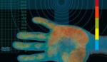
Non-ionizing medical imaging: Ultrasound, magnetic resonance imaging (MRI), macroscopic anatomy.
Ionizing medical imaging: Planar x-ray, computed tomography (CT), Positron emission tomography (PET).
The following aspects are covered by the course:
Operation and properties of the scanners. Hands-on experience with the scanners. Physical principles of the above imaging modalities. Contrast and resolution size. Image processing, analysis and visualization.
Grading: The grading in the course is based on:
- The report (40%).
- Written 2-hour multiple-choice exam (60%).
Format: Project work with concurrent lectures on the theoretical subjects, tools and methods. The students form teams of four. Each team is given their own phantom (depending on number of course participants), which will be scanned at a hospital with the imaging modalities covered by the course. Data is given to the teams for processing, analysis and visualization. Each team selects a scan plane and extracts the image at this plane for all imaging modalities and compares these (possibly very different) data. The phantom is finally sliced to produce a macroscopic anatomy picture for comparison with the aforementioned images.
Limitation in number of course participants: The students in the BSc of Biomedical Engineering (Medicin & Teknologi) will take about 50 seats, leaving the remaining seats for regular DTU students, external students and international students (see course description at menu item "Description" to the left).
| 


 Print Version